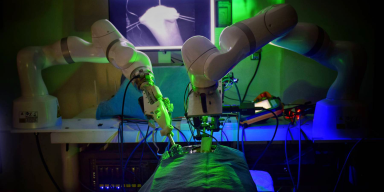
In 2016, a team at Johns Hopkins University (JHU), had demonstrated in a study that STAR, a surgical robot, could adapt to the subtle movement and deformation of soft tissue to perform precise and consistent sutures. Simon Leonard, an assistant research professor in JHU’s Whiting School of Engineering, had worked for four years to program the robotic arm. An enhanced version of STAR was developed to perform laparoscopy fully autonomously. Testing against manual laparoscopy and robotic-assisted surgery for porcine intestinal anastomosis, the researchers found that the autonomous surgery offered by the STAR system was more accurate and therefore superior.
Laparoscopy, more commonly known as laparoscopy, is performed by inserting an optical device and various instruments into the peritoneal cavity to view organs and perform a procedure without opening the abdomen. Surgeons are increasingly using this method, which allows the patient to recover much faster. Many scientists are trying to develop robotic assistants to help them during these operations such as Hominis, for example.
The team that designed STAR in 2016 started from the observation that even the surgeon’s safest hand is not as stable and consistent as a robotic arm built of metal and plastic, programmed to perform the same repetitive movements. She was able to demonstrate the precision and consistency of the robot, but the operation (intestinal anastomosis) required a large incision in the abdomen and supervision by the surgeon. Anastomosis, the suturing of two structures such as blood vessels or, in this study, two intestinal ends, is very delicate because the soft tissue can shift and change shape in complex ways while the stitching must continue. According to the researchers, complications such as leakage along the seams occur nearly 20 percent of the time in colorectal surgery and 25 to 30 percent of the time in abdominal surgery.
The new STAR
Autonomous anastomosis requires complex imaging, tissue tracking, and surgical planning techniques, as well as precise execution, often in unstructured and deformable environments; it is even more challenging when performed laparoscopically.
Researchers at Children’s National Hospital in Washington, D.C., including Axel Krieger, and Jin Kang, professor of electrical and computer engineering at Johns Hopkins, have enhanced the robot with new features for increased autonomy and surgical precision, using specialized suturing tools and advanced imaging systems that provide more accurate visualizations of the surgical field. A three-dimensional structural light-based endoscope and a machine learning-based tracking algorithm developed by Kang and his students guide STAR. Jin Kang states:
“We believe that an advanced three-dimensional computer vision system is essential to make intelligent surgical robots smarter and safer.”
In addition, an autonomous control system adapts the surgical plan in real time, just as a surgeon would. For his part, Axel Krieger adds:
“What makes STAR special is that it is the first robotic system to plan, adapt and execute a surgical plan in soft tissue with minimal human intervention.”
Study findings
Axel Krieger, lead author of the study, states:
“Our results show that we can automate one of the most complex and delicate tasks in surgery: reconnecting the two ends of a bowel. The STAR performed the procedure in four animals and produced significantly better results than humans performing the same procedure.”
Also according to him:
“robotic anastomosis will ensure that surgical tasks that require high precision and repeatability can be performed with greater accuracy and precision for each patient, regardless of surgeon skill. In our view, this will result in a democratization of surgical care with more predictable outcomes.”
Article source: autonomous robotic laparoscopic surgery for intestinal anastomosis
Translated from Robotique : le robot chirurgien STAR effectue des coelioscopies de façon autonome


