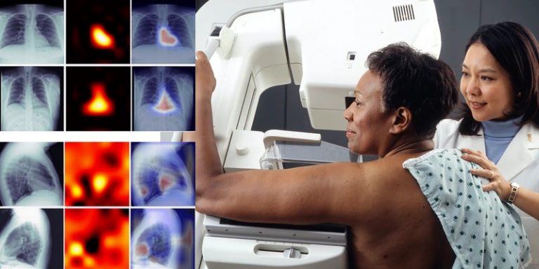
Several American radiologists and researchers have been working on deep learning to identify calcifications in the coronary arteries. Using an X-ray, it would be possible – by exploiting the capabilities of deep convolution neural networks – to assess this possibility. The model could then reduce cardiovascular risk by analyzing chest X-rays.
A study to quantify coronary calcifications on chest X-rays
In routine clinical practice, if a physician wishes to analyze an individual’s chest cavity, he or she has two options: either a CT scan or a chest X-ray. The latter is the more commonly used method, despite the fact that coronary artery calcifications (an abnormal accumulation of calcium deposits in this type of artery) are often overlooked during a medical check-up of the chest.
To help doctors analyze for this possibility, a team of radiologists affiliated with the Department of Radiology at Johns Hopkins School of Medicine have developed an innovative deep learning model. Their work was the subject ofa paper by Peter I. Kamel, Paul H. Yi, Haris I. Sair and Cheng Ting Lin.
Radiologists exploit deep learning and deep convolution neural networks
As part of their research, these radiologists have used recent advances in deep learning and deep convolution neural networks (DCNNs) to quantify information beyond what a human being can perceive. In order to be trained, the model designed by the researchers tapped into 1689 chest X-rays of patients who also received a cardiac scan in the same year, between 2013 and 2018.
The DCNNs were trained so that they could propose a binary classification on three aspects:
- Total calcium score: zero or non-zero.
- Calcium in each coronary artery: presence or absence.
- Total calcium score: above or below a variable threshold.
The results of this test image classification were compared to the 10-year arteriosclerotic cardiovascular disease risk scores in each cohort. By analyzing the area under the ROC (receiver operating characteristic) curve or AUC, the researchers were able to define the performance of the classifier.
Encouraging tests for further exploitation of deep learning in radiography
After analysis, the binary classification between zero and non-zero total calcium scores achieved an AUC of 0.73 on frontal chest X-rays, 0.70 for profile X-rays and 0.74 for the binary classification of total calcium score above or below a given value. Furthermore, the algorithm can predict a non-zero calcium score independently of traditional cardiovascular risk factors: a feature demonstrated using multivariate logistic regression.
From these results, the researchers stated that DCNN trained on chest X-rays had modest accuracy in predicting coronary artery calcium in relation to cardiovascular risk. Their findings can be used as a basis for future applications of deep learning in radiology to confirm these results or to improve them.
Translated from Des radiologues s’intéressent au deep learning pour diagnostiquer les calcifications coronaires


