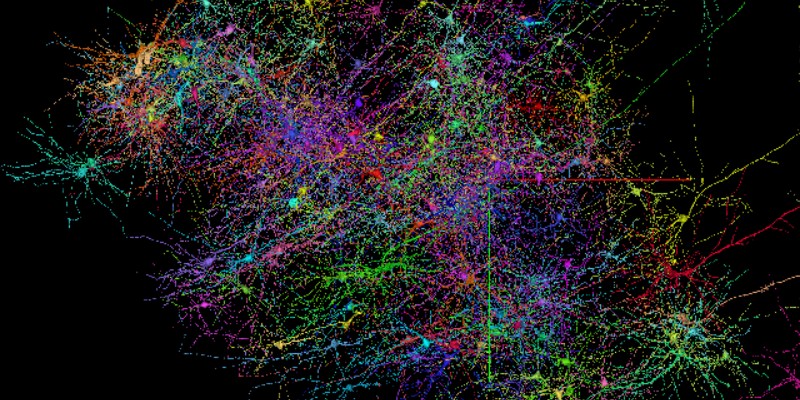
At the beginning of June, Google announced, in collaboration with the Lichtman laboratory of Harvard University, the realization of a cartography of the human cerebral cortex in three dimensions. In parallel, a 1.4 petabyte (1015bytes) dataset has been published: it includes imaging data covering about 1 mm3 of brain tissue as well as tens of thousands of reconstructed neurons. This tool should help advance studies of neural networks in the human brain, which are currently difficult.
Google AI and Harvard University researchers map the cerebral cortex in 3D
Thanks to a collaboration between Harvard University's Lichtman Laboratory and Google AI, researchers have successfully mapped the human cerebral cortex in 3D. Already in January 2020, Google offered a database providing information about the morphological structure and synaptic connectivity of half of a fly's brain. From this research around the brain of flies, the research teams then turned to the human brain.The cerebral cortex (or grey matter) is the thin superficial layer found in vertebrate species. In mammals, and especially in humans, it plays an important role in the individual's head. It plays a crucial role in most higher-level cognitive functions, such as thinking, memory, planning, perception, language and attention.
Although there has been some progress in understanding its macroscopic organization, little is known about its organization at the level of nerve cells and interconnected synapses. It is to help research on these two aspects that researchers decided to design a 3D map of the human cerebral cortex.
A data set of nearly 1.4 petabytes
The "HO1" dataset is a 1.4 petabyte data set of a small sample of human brain tissue accompanied by a paper entitled "A connectomic study of a petascale fragment of human cerebral cortex" whose lead author is Alexander Shapson-Coe, from the Department of Cellular and Molecular Biology at Harvard University, and which shows how this database has been exploited to study the cerebral cortex.It includes imaging data that covers about a cubic millimeter of brain tissue and includes tens of thousands of reconstructed neurons, millions of neuron fragments, 130 million annotated synapses, 104 reread cells and many additional annotations and subcellular structures. The "H01" sample was imaged at 4 nm resolution by electron microscopy, reconstructed and annotated by automated computational techniques, and analyzed to obtain preliminary information on the structure of the human cortex.
The 3D mapping and database are freely available online. In the future, researchers will face the problem of data storage if they wish to continue their work and model the rest of the human cerebral cortex in 3D. For example, the entire brain of a mouse could generate nearly one exabyte (1018 bytes) of data.
Translated from Google AI et l'Université de Harvard cartographient une partie du cortex cérébral en trois dimensions


