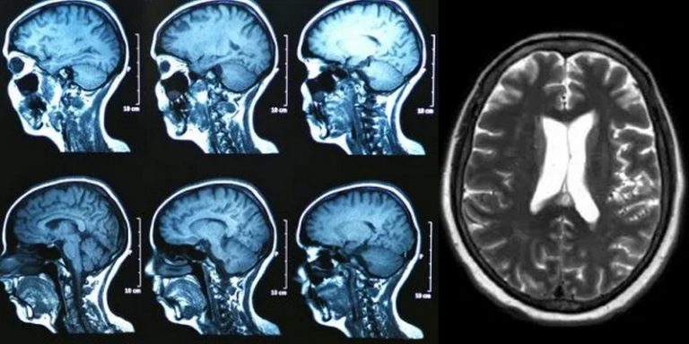
To classify a brain tumour into one of six common types using a single 3D MRI scan: that was the challenge set by a team of American researchers. Together, they designed a deep learning model capable of this feat, a step closer to creating a reliable tool for automatic detection of tumors and the absence of tumor pathology images. The experts used a large intracranial 3D MRI dataset to train and test the system.
The challenge of automating brain tumor detection using 3D MRI
To succeed in treating the most common intracranial tumors more easily, to determine directly the tumor class or its absence with the help of a 3D MRI, and to adapt the follow-up and treatment of the patient according to this classification. That’s the challenge facing the Digital Imaging Laboratory at the Mallinckrodt Institute of Radiology at Washington University School of Medicine in Saint Louis, Missouri.
Satrajit Chakrabarty, a doctoral student under the supervision of Prof. Aristeidis Sotiras, and Prof. Daniel Marcus with the help of Pamela LaMontagne, Michael Hileman and Daniel Marcus have written a paper around the deep learning model they designed that is able to classify a brain tumor into one of six common types, using a single 3D MRI, according to the study.
High-grade glioma, low-grade glioma, brain metastasis, meningioma, pituitary adenoma, and acoustic neuroma: these are the six most common types of intercranial tumors, and each of them requires surgical removal of the suspected cancerous area and examination under a microscope.
Deep learning and convolutional neural networks
To design their deep learning model, the researchers developed a large multi-institutional intracranial 3D MRI dataset from four publicly available sources. In addition, the team obtained preoperative MRI scans from the segmentation of known brain tumor images.
In total, 2,105 scans were divided into three subsets of data:
- 1396 examinations were used to train the model, specifically to train the convolutional neural network to distinguish between healthy images and tumor identifications, and to classify tumors by type.
- 361 reviews for internal testing and 348 for external testing. These data were used to evaluate the performance of the model.
Very encouraging results for the classification of intracranial tumors
With the in-house test data, the model achieved an accuracy of 93.35% (337 out of 361) in seven image groups (one healthy and six tumor classes). Sensitivities ranged from 91% to 100%, and positive predictive value ranged from 85% to 100%. Negative predictive values ranged from 98% to 100% in all classes. With the external testing data (which is only for high-grade glioma and low-grade glioma), the model is 91.95% accurate.
The researchers consider that with these results, deep learning is a promising approach for the automated classification and assessment of brain tumors. They add that the use of convolutional neural networks avoids the time-consuming step of tumor segmentation with classification. For Satrajit Chakrabaty, this research is just the first step towards an AI-enhanced radiology workflow.
Translated from Un modèle de deep learning permet de classer avec précision six types de tumeurs intracrâniennes


