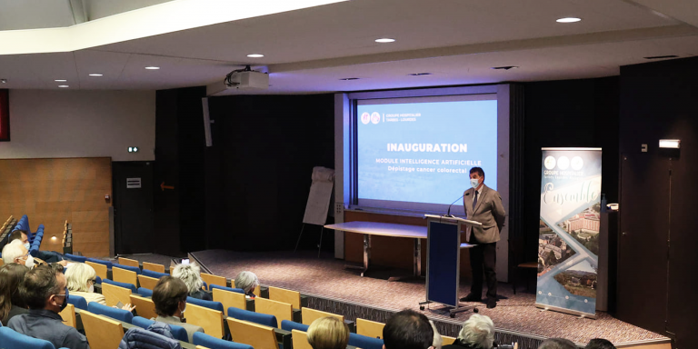
In mid-February, the Centre Hospitalier de Bigorre in Tarbes (Hautes-Pyrénées) organized the inauguration of an Artificial Intelligence module for digestive endoscopy, in order to optimize colorectal cancer screening: CAD EYE by Fujifilm. The hospital’s endoscopy department has already been using it for a year and is witnessing the benefits of such a technological innovation for the care of patients in the Hautes Pyrénées.
Colorectal cancer is the 3rd most common cancer after lung cancer and breast cancer, and the second most common cause of death by cancer after lung cancer. However, if detected at an early stage, colorectal cancer can be cured in 9 out of 10 cases, which is possible with colonoscopy (lower digestive endoscopy) for the detection of colon tumors. On the other hand, an accurate endoscopic diagnosis of colon polyps could reduce the number of unnecessary polypectomies.
In March 2021, the endoscopy department was able to acquire the Fujifilm CAD EYE box equipped with artificial intelligence thanks to the financing and the important mobilization of the League against Cancer of the Hautes Pyrénées, the Departmental Council, the Lions Club and the Rotary Club, organizers of the Maxi Loto of Lourdes, for the benefit of cancer research. Annette CUQ, President of the CD65 of the League against cancer, declares:
“With the current vice-presidents and Dr. Bernard COUDERC (oncologist and former President of the Departmental Committee of the League), we were very quickly convinced by the presentation of Dr. ANDRAU and wanted to quickly mobilize our personal knowledge to obtain this funding. This project seems to us to be of primary importance and interest given the scientific progress that it represents for screening, both for the population of the Hautes Pyrénées and for the work of endoscopists. We were delighted to get such a strong support from all the partners in such a short time.
The CAD EYE AI system
CAD EYE provides real-time detection of colon polyps using AI technology. When a suspicious polyp is detected in the endoscopic image, a detection box indicates the area where the suspicious polyp was detected, and an audible signal is heard. The CAD EYE characterization module supports clinicians by generating a histological prediction suggestion that specifies whether the suspected polyp in the image is hyperplastic or neoplastic.
Fujifilm had previously developed two different image enhancement technologies called LCI (Linked Color Imaging) and BLI (Blue Light Imaging) to support detection and characterization, respectively, through the different wavelengths of light used. Fujifilm has integrated these technologies into the development of CAD EYE, whose features are automatically activated depending on the observation mode used.
The display of the characterization result appears just below the endoscopic image as well as a visual assistance circle placed on its circumference. Finally, the position map is placed directly next to the clinical image to show which part of the video image CAD EYE is focusing on. Professor Helmut Neumann, Professor of Medicine and Director of the Interdisciplinary Center for Endoscopy at the University Medical Center Mainz, states:
“Increasing the number of endoscopists who are able to correctly detect and characterize colonic polyps is one of the critical issues in the field of gastroenterology. With the combination of detection and characterization offered with CAD EYE, the learning curve for colonoscopy exams can be greatly improved. With the help of CAD EYE Detection, we may see an increase in polyp detection rates, and even those who are not endoscopy experts can get much closer to that level. CAD EYE Characterization can reduce the cost of histopathology by reducing the number of unnecessary biopsies taken during endoscopies.”
Translated from L’Intelligence Artificielle au service du dépistage du cancer colorectal


